Researchers simulated a living cell in 3D thanks to the power of GPUs
Every living cell has a “microcosm“made up of thousands of components responsible for energy production, protein construction, gene transcription and more. Understand how all components interact and change in response to internal and external signals helps to better understand the fundamentals of life, this is why the simulation carried out by a team from the University of Illinois at Urbana-Champaign relying on GPUs, in this case those of NVIDIA, is important.
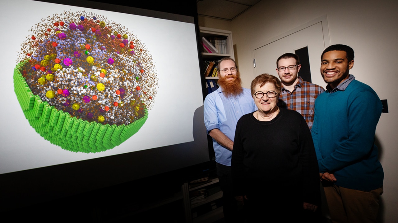
The group of scientists is succeeded to recreate a 3D simulation that replicates to scale the physical and chemical characteristics of a “minimal” living cell, developing a fully dynamic model that mimics its behavior. The study was published in the Cell.
The minimal cell contains a reduced set of genes essential for survival, functioning and replication. These cells are simpler than natural ones, which makes them easier to recreate digitally. “Even a tiny cell requires 2 billion atoms,” said Zaida Luthey-Schulten, professor of chemistry and co-director of the university’s Center for the Physics of Living Cells. “You can’t make a 3D model like this on a realistic human time scale without the GPUs“.
The model is based on the use of NVIDIA GPUs for simulate 7000 processes of genetic information over a 20 minute span of the cell cycle, making it what scientists believe is the longest and most complex cell simulation to date. Once further tested and refined, the complete cell models can help scientists predict how changes to real-world cell conditions or genomes will affect their function.
To build the living cell model, the Illinois researchers have simulated one of the simplest living cells, a parasitic bacterium called mycoplasma. They based the model on a scaled-down version of a mycoplasma cell synthesized by scientists at the J. Craig Venter Institute in La Jolla, California, which was just under 500 genes to keep it viable. For comparison, a single E. coli cell has around 5,000 genes. A human cell has more than 20,000.
Luthy-Schulten’s team then used known properties of the inner workings of mycoplasma, including amino acids, nucleotides, lipids, and small molecule metabolites to build the model with DNA, RNA, proteins and membranes. “We had enough reactions to reproduce everything we knew,” he said.
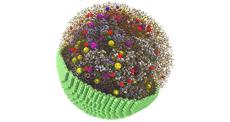
Snapshot of the 20-minute 3D space simulation, showing yellow and purple ribosomes, red and blue degasomes, and smaller spheres representing DNA polymers and proteins.
Through the Lattice Microbes software capable of harnessing the tensor cores of NVIDIA GPUs, the researchers ran a 20-minute 3D simulation of the cell’s life cycle, before it begins to substantially expand or replicate its DNA. The model showed that the cell he devoted most of his energy to the transport of molecules across the cell membrane, which fits the profile of a parasite cell that gets most of what it needs to survive from other organisms.
“If I performed these calculations in series, or at the level of all atoms, it would take years“said Zane Thornburg, lead author of the study.” But because they are all independent processes, we can bring parallelization into the code and use GPUs. “
The simulations also allowed Thornburg to calculate the natural lifespan of messenger RNAs, the genetic blueprints for building proteins. They also revealed a relationship between the rate at which membrane lipids and proteins were synthesized and changes in the membrane surface area and cell volume.
“We simulated all the chemical reactions within a minimal cell, from its birth until the time it divides two hours later,” Thornburg said. ‘From this, we get a model that tells us how the cell behaves and how we can make it more complex to change its behavior,’ said Professor Luthey-Schulten.
Thornburg is working on another GPU-accelerated project to simulate cell growth and division in 3D. “The team recently adopted NVIDIA DGX systems and RTX A5000 GPUs to accelerate their work,” notes NVIDIA, “noting that using A5000 GPUs improved simulation time by 40% compared to a workstation with a GPU. Previous Generation NVIDIA “.
Related posts:
 7-nanometer Nvidia GPU, TSMC will handle most of the production
  ASRock X299, a new BIOS allows you to install 2 TB of memory
  MSI Prestige X570 Creation Review: Test | Specs | Hashrate
  Radeon RX 5500 XT, PCI Express 3.0 castrates performance?
  An overclocker ran 1TB of RAM on an X299 motherboard limited to 256GB
  Best Review 2021: MSI MPG X570 Gaming Edge WiFi Under $250 ($200)